

Therapy of aortic aneurysm diseases
The most common disease of the aorta is an enlargement (aneurysm). An aortic aneurysm is an enlargement of the aorta by more than twice the usual diameter of 2 to 3 cm.
Depending on where the dilation is located, either an endovascular stent or a surgical vessel replacement is used to eliminate the abnormal dilation of the aorta and prevent the life-threatening tearing of the aortic wall (aortic dissection).
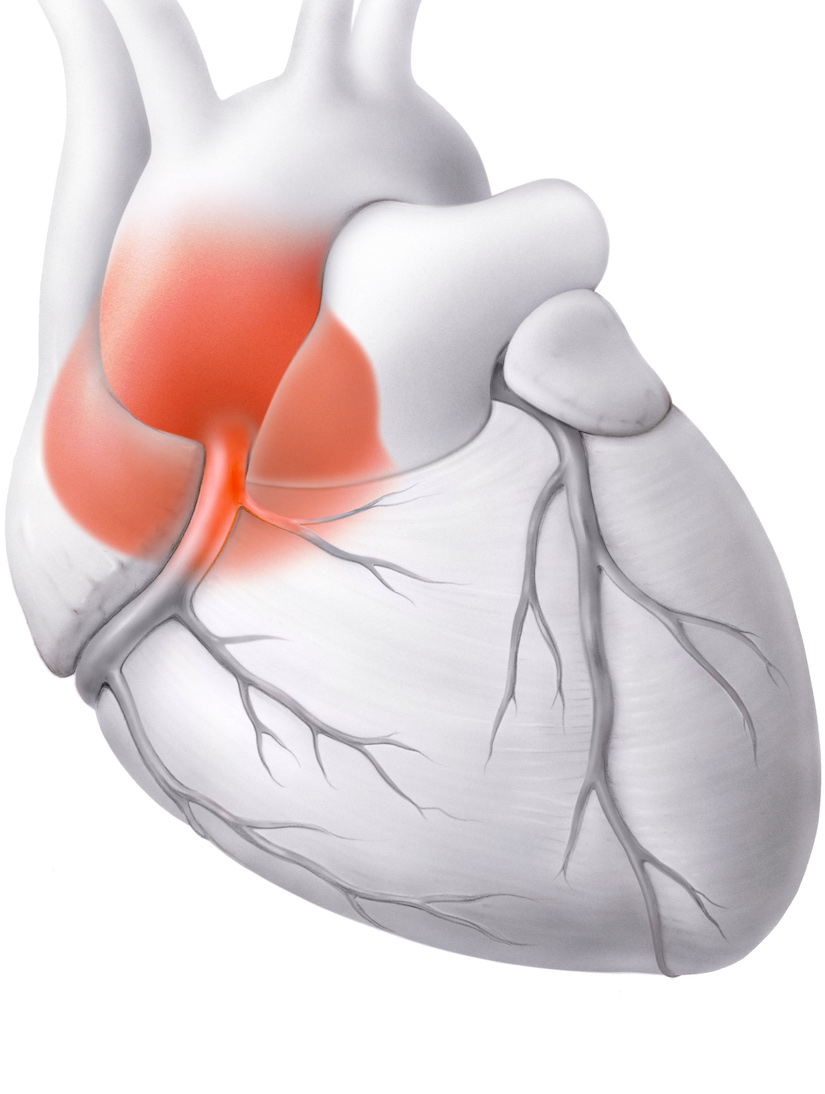
Ascending aorta as a particular danger
Dilation of the ascending aorta (ascending aorta close to the heart) is particularly dangerous, as it can often rupture or split at a diameter of just 5 cm. Elective (timely planned) replacement of the ascending aorta is an established and effective prophylaxis.
In any case, surgery is indicated if the diameter of the vessel in an adult exceeds 5.5 cm (in Marfan syndrome or other connective tissue diseases from 5 cm).
Target groups and causes of aortic aneurysm
Aortic aneurysms occur most frequently between the ages of 60 and 70, with men being affected two to four times as often as women. The cause is usually advanced hardening of the arteries (atherosclerosis) resulting in high blood pressure that is not controlled by medication.
Smoking and old age also favor the formation of an aortic aneurysm.
Screening and follow-up of aortic aneurysm
The method of choice for screening and, if necessary, for follow-up checks is transthoracic echocardiography (cardiac ultrasound), or TTE for short. The maximum vessel diameter of the aorta is decisive for diagnosis and treatment.
In the case of echocardiographic evidence of a dilated aorta or poor visualization, our specialists use computed tomography (CT) or magnetic resonance imaging (MRI) to assess the full extent of the dilatation.
Preparations before an aortic aneurysm intervention
Before an aortic aneurysm intervention, the exact location and extent of the aneurysm is determined using ultrasound, computed tomography (CT) or magnetic resonance imaging (MRI).
Above and below the aneurysm, there must be a sufficiently long piece of non-dilated aortic wall without major vascular outlets so that a vascular prosthesis can be inserted with good support.
As arteriosclerosis of the aorta often also affects the coronary or cerebral arteries, these vessels are also examined in detail before surgery. Constrictions of the coronary or cerebral arteries may need to be repaired before or, in the case of open surgery, in combination with aortic intervention in order to minimize the risk of a heart attack or cerebral infarction occurring during or after the procedure.
In preparation for the operation, the usual pre-operative examinations are also carried out (ECG, blood pressure measurement and blood test). Strong blood thinners must be discontinued before the operation.
Our therapies for aortic aneurysm diseases


Your contacts for aortic aneurysm


Heart Surgery